Early access: pre-order PoroScan Academic now. Deliveries start in December 2026, after CE marking. Pre-order now
PoroScan Academic
See bone quality DXA can’t.
PoroScan Academic measures the microstructure of cortical bone with ultrasound: radiation-free, in about ten minutes, and sensitive to change within months.
Invented at Charité – Universitätsmedizin Berlin
1,600-participant multicentre study
ISO 13485 certified quality management
Market entry December 2026, pre-orders open
What PoroScan Academic adds to your bone research.
DXA tells you how dense bone is. HR-pQCT shows structure, but only at a few sites and with radiation. PoroScan Academic brings cortical microstructure to your own site, radiation-free.
DXA
HR-pQCT
PoroScan Academic
What is measured
DXA
Areal bone mineral density
HR-pQCT
3D bone microstructure
PoroScan Academic
Cortical microstructure and viscoelasticity: thickness, sound velocity, attenuation, pore-size distribution, porosity
Ionising radiation
DXA
Yes (low dose)
HR-pQCT
Yes (low dose)
PoroScan Academic
None, ultrasound only
Repeat measurements
DXA
Limited by dose
HR-pQCT
Limited by dose
PoroScan Academic
As often as your protocol needs
Children, women of childbearing age
DXA
Restricted
HR-pQCT
Restricted
PoroScan Academic
Possible
Where it runs
DXA
Imaging centre
HR-pQCT
Few specialised centres
PoroScan Academic
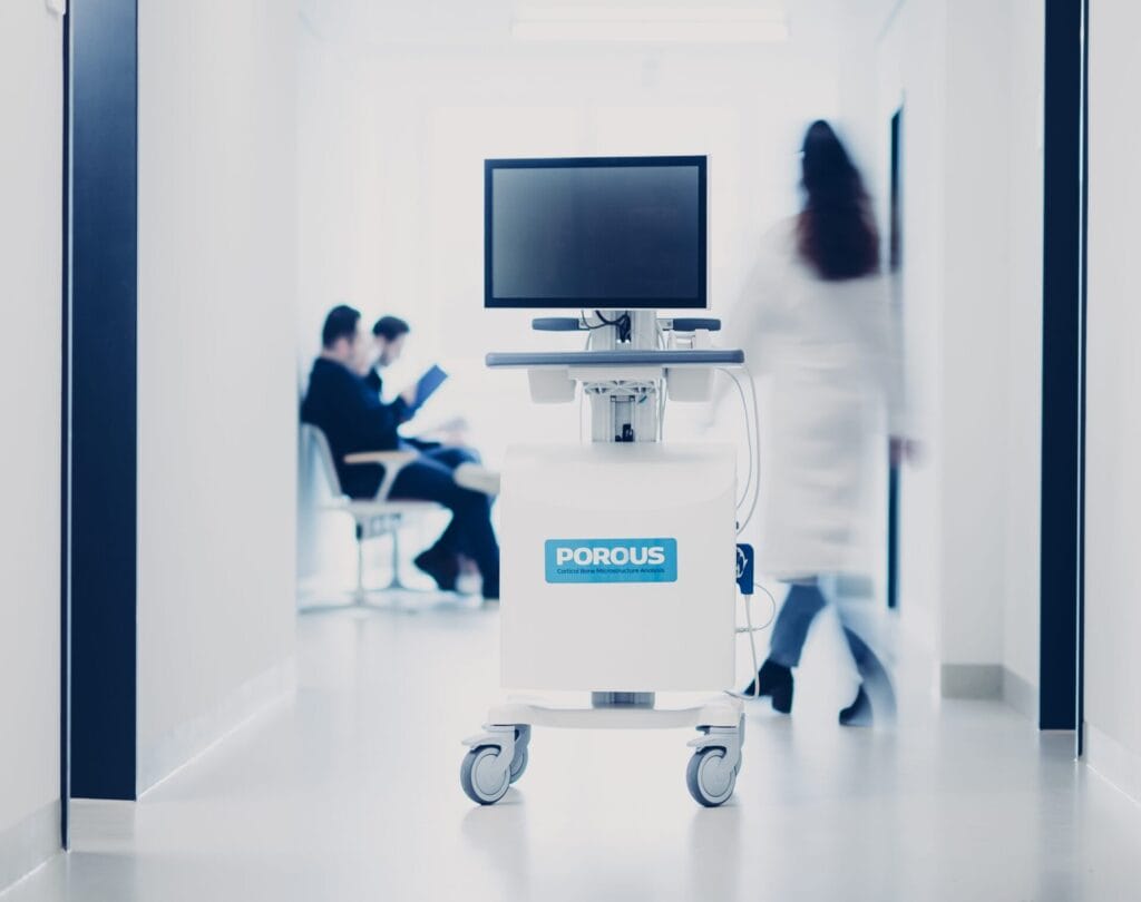
Mobile cart at your site
Change visible after
DXA
12–18 months
HR-pQCT
Not established
PoroScan Academic
Months¹
¹ Sensitive to short-term changes, shown in bed rest and exercise countermeasures: in the ESA/CNES BRACE bed-rest study (MEDES, Toulouse), analysed by Charité – Universitätsmedizin Berlin, the width of the pore-size distribution increased by 35 % and mean pore size by 15 % after 60 days of bed rest without exercise, while cycling training prevented these changes (Raum et al., Osteologie 2025). Data acquired with the predecessor system R1C, which uses the same measurement principle. PoroScan Academic complements established reference methods; it does not replace them.
Three steps. About ten minutes.
1
Position
The participant sits or lies with the leg resting. The probe is placed at mid-shin, and a live ultrasound image guides the operator to the bone surface.
2
Scan
A guided 3D sweep with the motion-tracked probe captures the volume of interest. No radiation, no special room.
3
Analyse
Fully automatic processing in under five minutes delivers cortical thickness, sound velocity, attenuation, pore-size distribution and porosity, plus the raw data.
Operated by trained staff after POROUS product training. No physician time required.
Built for bone research and clinical trials.
For research teams
New biomarkers. New publications. No radiation.
- Cortical microstructure that so far required high-resolution CT, on a mobile cart at your own site.
- More than 15 parameters per measurement: thickness, radial sound velocity, attenuation, backscatter and pore-size distribution.
- Measure as often as your protocol needs, including children, young adults and longitudinal designs.
- Standardised, quality-controlled acquisition for comparable data across sites and operators.
- Full data access, plus support for your grant application and study design.
Applications: bone metabolism, bed rest and space medicine, osteoporosis and osteopenia, secondary osteoporosis, rare bone diseases, paediatrics, nutrition and lifestyle, animal science.
For pharma and clinical trials
See treatment effects in months, not years.
- Follow bone changes at short intervals instead of waiting 12–18 months for DXA.
- Build evidence of treatment effectiveness early, for development decisions and payer discussions.
- Regular, radiation-free follow-ups that can support patients in staying on therapy.
- Multi-parameter readout with the potential for treatment-specific monitoring.
- A complete pilot programme at your study sites: devices, operator training, data management and analysis.
Built on clinical evidence.
~2,000
participants measured in POROUS clinical studies
In a Charité pilot study with 55 postmenopausal women, biomarkers measured with the POROUS method discriminated fragility fractures as well as high-resolution CT and better than DXA (Armbrecht et al., JBMR Plus 2021). All publications
Choose how you want to start.
Early access
Pre-order your system
Reserve one of the first PoroScan Academic systems, including software licence, operator training, service and maintenance. Deliveries start in December 2026, after CE marking.
Pilot study programme
For pharma and industry: we deliver devices, operator training, data management and analysis at your study sites.
Scanning and analysis service
We measure and analyse for you, and support your study design and grant application.
Device
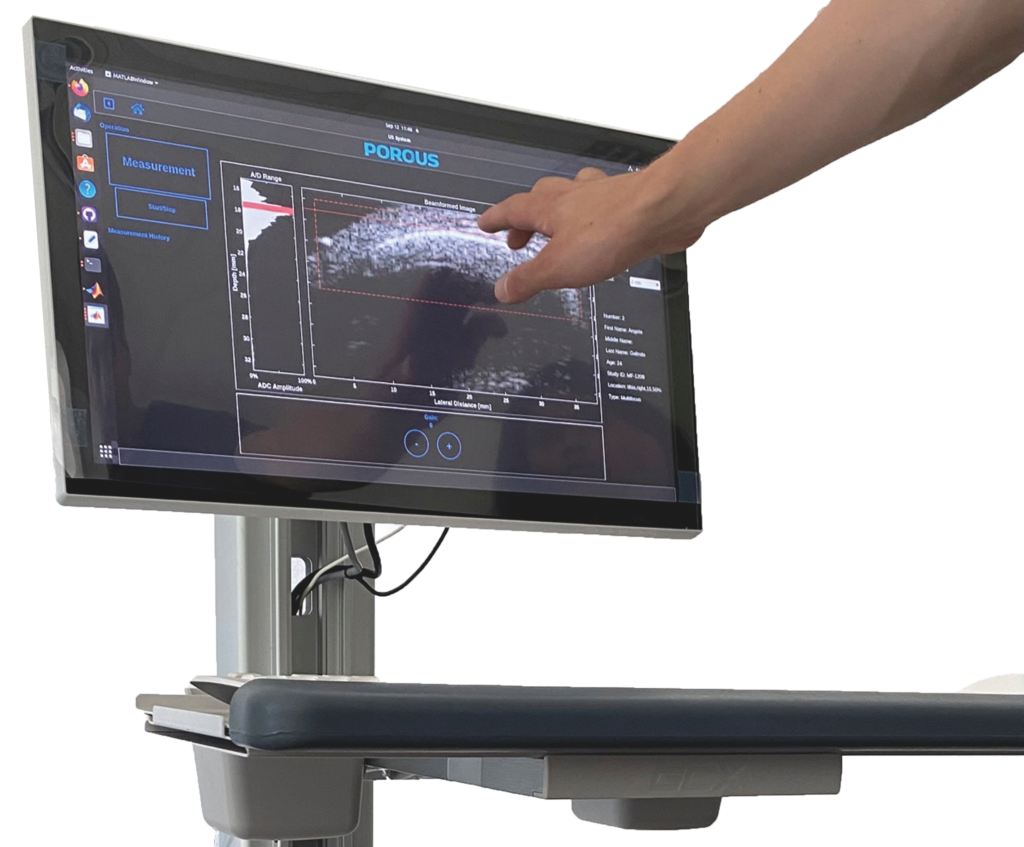
PoroScan Academic (POROUS R2C): mobile cart system with 21″ touch screen and guided 3D probe
Measurement site
Tibia (mid-shin)
Biomarkers
Cortical thickness, sound velocity, attenuation, backscatter, pore-size distribution, porosity
Measurement time
About 10 minutes including automatic analysis
Radiation
None, ultrasound only
Data
Report per measurement, local database
Dimensions and weight
83 × 80 × 130 cm, 55 kg
Regulatory
Class IIa medical device (MDR). CE marking pending, expected December 2026
PoroScan Academic is not yet CE-marked and will be delivered only after CE marking. Intended for the investigation and assessment of cortical bone state and quality by trained personnel; not intended for diagnostic use.
PoroScan in action
1,500+
pig bones measured and analysed so far
PigBoneS: healthier, more sustainable livestock
Challenge: improve phosphorus utilisation and bone stability in pigs while reducing feed additives and emissions.
Approach: Eurostars project with SUISAG and FBN, bringing non-invasive ultrasound bone assessment into modern breeding.
Status: project ongoing, results pending.


60 days
of bed rest: changes in cortical pore structure and viscoelasticity detected
ESA/CNES BRACE bed-rest study
Challenge: track changes in cortical bone during immobilisation, for space medicine and rehabilitation.
Approach: tibia ultrasound in the ESA/CNES BRACE bed-rest study (MEDES, Toulouse), analysed by Charité – Universitätsmedizin Berlin. Data acquired with the predecessor system R1C, which uses the same measurement principle.
Result: without exercise, mean pore size increased by 15 % and the width of the pore-size distribution by 35 %, and viscoelastic attenuation decreased. Cycling training prevented these changes; artificial gravity added no further benefit (Raum et al., Osteologie 2025).
Frequently asked questions
Can’t find your answer? Contact us
Does PoroScan measure bone mineral density?
No. PoroScan does not measure bone mineral density like DXA. It quantifies the structure and acoustic properties of cortical bone, information that complements density measurements.
How difficult is it to take measurements?
The participant sits or lies with the leg resting. The operator places the probe at mid-shin, aligns the bone surface with the help of a live ultrasound image and sweeps the probe with visual guidance. All analysis steps are automatic.
How long does a measurement take?
About ten minutes including positioning. The automatic data processing takes under five minutes.
Does the measurement use radiation?
No. PoroScan uses ultrasound only, so measurements can be repeated as often as needed, also in children and young adults.
Who can operate the device?
Healthcare professionals and trained research staff who have completed the POROUS product training. No physician is required to perform the measurement.
Why do you measure at the tibia?
The tibia is a weight-bearing bone with a thick cortex directly under the skin, which allows clear ultrasound access. Measurements are standardised at 50 % of the tibia length.
How does PoroScan differ from older bone ultrasound devices?
Conventional bone ultrasound mostly tries to estimate bone density and mimic DXA results. PoroScan analyses the ultrasound signal scattered inside the cortical bone to quantify its microstructure, the properties that determine bone strength.
What exactly does PoroScan measure?
Cortical thickness, radial speed of sound, frequency-dependent attenuation, backscatter coefficients, the distribution of cortical pore diameters and cortical porosity. Pore-related parameters are model-derived biomarkers, not direct images of individual pores.
Ready to add cortical bone biomarkers to your next study?
Tell us about your project. We will get back to you within two working days.
POROUS GmbH, Am Mühlenberg 11, 14476 Potsdam, Germany
+49 331 74899955, info@porous.care
PoroScan Academic (POROUS R2C) is intended for the investigation and assessment of cortical bone state and quality by trained personnel. It is not intended for diagnostic use and does not replace established reference methods. CE marking pending (expected December 2026); deliveries only after CE marking.